Gastrointestinal
The Gastrointestinal (GI) tract includes the mouth, oesophagus, stomach, liver, pancreas, small and large intestines. When diagnosing gastrointestinal disorders, accurate and precise diagnosis is crucial.
What is GI Histology?
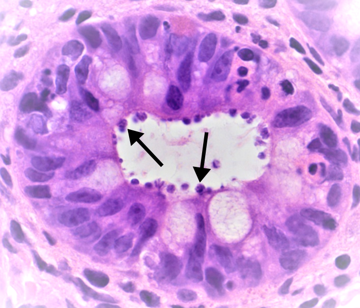
Histopathology is the science of examining cells and tissues from biopsies or surgical operations in order to establish a diagnosis to find therapy options. We work with consultants and hospitals to provide further testing and advice by analysing biopsies from endoscopies and operations to diagnose many other GI diseases. For instance, the photo below shows organisms in the glands of the colon which can only be identified by histology and is effectively treated by antibiotics.
Recent advances in medicine allow us to deploy techniques such as immunohistochemistry (using antibodies with a visible “tag” to localise target molecules in tissues) or “molecular analysis” which can involve extracting DNA and RNA from the tissues to see what genes may be altered and which then serve as targets for the best treatments for cancer and other gastrointestinal disorders.
Image (1) is an example; Some gastric cancers can over-produce a protein called HER2 due to a cancer-specific change in the DNA. We can see the brown dye ‘tag’ sticking to the cell membranes very strongly. This means the tumour may respond to drugs well established in the treatment of breast cancers and they can be very effective in otherwise hard to treat cases.
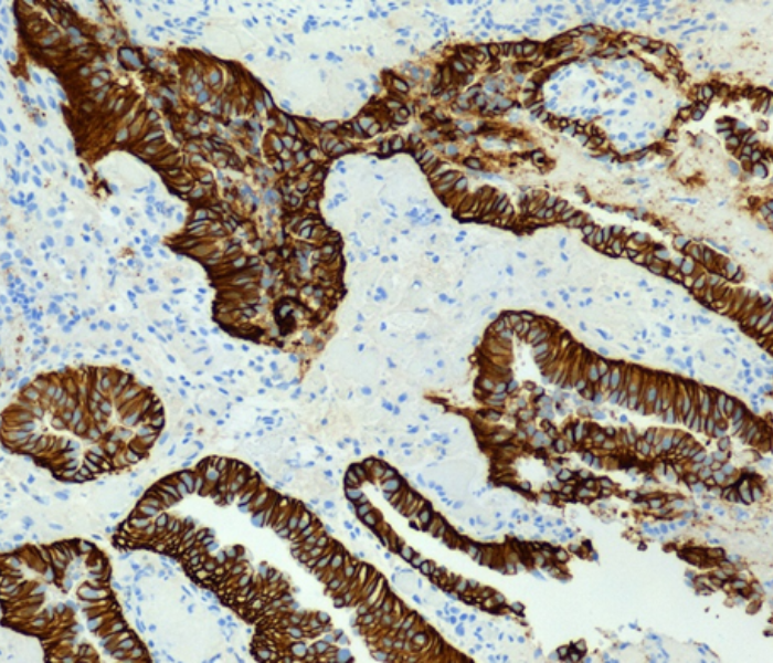
Image (1)
GI Histology at AHL
1/ Accurate diagnosis
At AHLab our team of highly experienced consultant histopathologists include Dr Patrizia Cohen, Dr Vinu Sheshappanavar, Prof Gordon Stamp and Dr Morgan Moorghen, who specialise in diagnosing conditions of the GI tract. Their expert opinions facilitate personalised patient treatment due to their level of knowledge in specific GI disorders, distinguish between different subtypes, and differentiate between mimicking conditions.
Image (2) is a biopsy from a patient with very severe heartburn and swallowing difficulties. It demonstrates a condition called eosinophilic oesophagitis, which is triggered by some allergens in food. In the image you can see eosinophils which have bright red granules in the cytoplasm, containing histamine and other chemicals the use to deal with allergic reactions. These are causing fluid to build up in the lining epithelium resulting in the white spaces and separation of the cells which are normally a barrier. Once diagnosed, this can be treated by expert gastroenterologists.
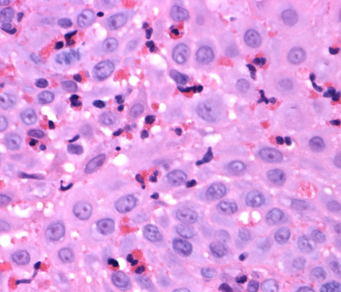
Image (2)
2/ Early detection of GI diseases
As with most diseases early detection is crucial to achieve positive patient outcomes. GI histology assists with the early detection of gastrointestinal disease by identifying any abnormal cellular changes, usually prior to any clinical symptoms arising.
It may seem strange now but 40 years ago it was not uncommon to hear of people having “stomach ulcers” that in some cases led to death from haemorrhage and peritonitis when they burst. Today, this is very uncommon and it is due to the determination of a pathologist Robin Warren and researcher Barry Marshall, in Australia, who demonstrated there were bacteria, Helicobacter pylori, that lived in the stomach which caused gastritis and most ulcers, not stress as was commonly believed. Moreover H pylori are linked to the development of gastric cancers and some rare lymphomas. These days, these organisms and the effects they are having can be recognised on biopsies. Image (3) shows a simple stain that we use, Cresyl Fast Violet, which will stain the bacteria allowing their detection.
They can be eliminated by a course of antibiotics, so these days not only are gastric ulcers rare, there is considerable social benefit from reducing gastritis. The process of eradicating H. pylori is now having benefits in reducing gastric cancer. An accessible account of this fascinating story can be found on the Guts UK website.
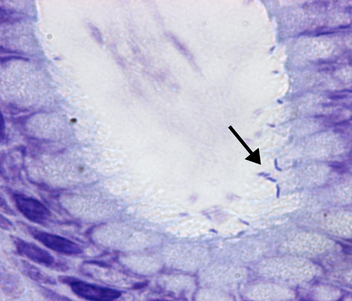
Image (3)
3/ Effective management of GI diseases
According to figures from Bowel Cancer UK, almost 43,000 people are diagnosed with bowel cancer every year in the UK but thankfully the number of people dying of bowel cancer has fallen since the 1970s. This reduction is in part due to factors such as earlier diagnosis and better treatment options for conditions predisposing to cancer development offered by GI histology. This minimises the need for unnecessary surgical procedures and treatments and offers more effective patient management including lifestyle changes.
Image (4) shows a typical cancer of the bowel which is invading into the pink staining muscle at the bottom of the field. Our pathologists assess how far the tumour has spread into the wall of the bowel which affects each stage and choice of future treatment.
Image (5) is the higher power image of the tumour which shows very disorganised glands which have caused some inflammatory reaction around them. We can extract DNA and RNA from this tissue to find out what mutations are present or attach antibodies to these sections to determine choices of treatment which will work best for the patients.
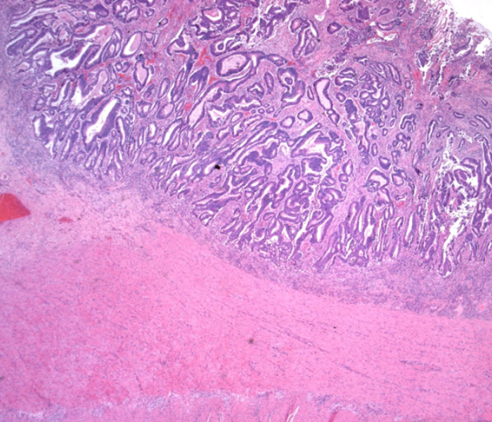
Image (4)
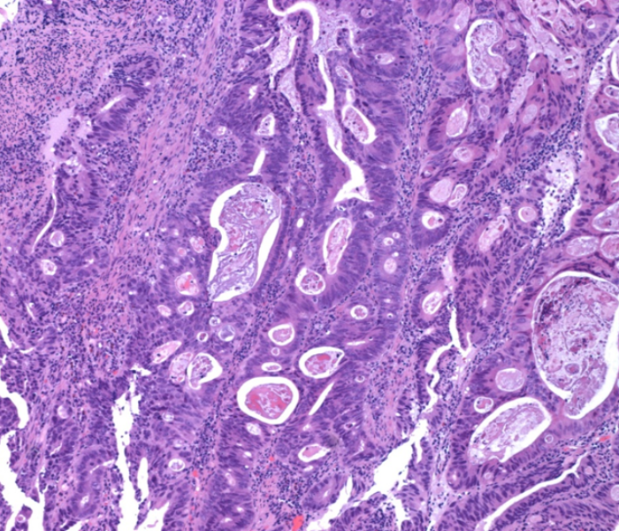
Image (5)
4/ Ongoing patient treatment
Once diagnosed, the ongoing management of GI diseases is an important part of effective ongoing patient treatment. Our team of consultant pathologists can periodically examine tissue samples to assess patient responses to therapy and treatments. This allows the identification of any treatment plans that may need to be modified to ensure the maximum benefit to patients.
At times patients and clinicians may seek reassurance about the original diagnosis formed elsewhere. At AHLab our Expert 2nd Opinions service provides access to an extensive referral network allowing us to confirm the initial diagnosis or suggest additional diagnostic procedures where this is required. Of paramount importance to us is the effective diagnosis to ensure the best possible patient treatment.
5/ Access to the latest scientific advances
One of the key benefits of GI histology is the vital role it plays in medical research and the development of new treatments. Studying GI diseases at a molecular level allows a greater understanding of the cellular changes that occur, this in turn leads to the development of innovative treatment options being developed.
AHLab Director and Consultant Histopathologist, Professor Gordon Stamp, is a key advocate of the importance of molecular analysis in providing access to the latest treatment options. He is an internationally renowned diagnostic pathologist with specific expertise in the ongoing development of oncology diagnosis.
Why choose AHLab?
Our high-quality & rapid GI histology diagnostic service provides clinicians with access to a wide network of expert consultant pathologists and scientists, experienced in diagnostic and research pathology, working in centres which are internationally recognised.
Utilising the latest technology and specialised equipment we offer rapid sample turnaround, high levels of expertise for in-depth analysis, a personalised service delivered by a contactable and accountable team of specialists, together with a competitive pricing structure.
Get in touch:
For more information about our GI histology diagnostic services, please read our user guide or contact us.
Email: [email protected]
Call: 020 7636 9447

